Contents
- a) What is advanced electrolysis blemish removal?
- b) Non-treatable & treatable conditions for electrolysis blemish removal.
- c) Casual factors that could cause skin lesions.
- d) Telangiectasia characteristics & causes.
- e) Spider naeivi characteristics & causes.
- f) Cherry angiomas aka Campbell de Morgan spots or blood spots characteristics & causes.
- g) Thread veins on the legs characteristics & causes.
- h) Skin tags characteristics & causes.
- i) Milia characteristics & treatment.
- j) Seborrhoeic warts or keratosis characteristics & causes.
- k) What is cryotherapy and how is it used to treat blemishes?
- l) Advantages and disadvantages of Cryopen.

Find Blemish Removal Treatments in Your Area

a) What is advanced electrolysis blemish removal?
Non-Surgical Blemish Removal is a very safe, effective, and quick procedure involving the removal of Telangiectasia (red veins), Spider Naevi, Skin Tags, Blood Spots, Seborrheic Keratosis, Milia, Hairy Moles and Viral Warts using electrical currents. It is commonly known as advanced electrolysis and has traditionally been carried out by beauty therapists who had initially qualified in electrolysis.
Due to the rapid growth of the clinical aesthetic industry these treatment are now carried out by aesthetic practitioners, doctors, nurses, dentists or those with other suitable clinical backgrounds.
Does it hurt?
A very fine needle probe is used to pass the heat of the electrical current into the tissue. This is so fine and localised that only the smallest amount of discomfort is experienced and for most people there is very little discomfort.
What are the side effects?
During the first few days the area will be sensitive, show redness and a little swelling. Larger lesions may develop scabbing. Providing the correct aftercare instruction is followed preventing infection, the risks of any scarring is minimal.
Deeper lesions may leave a faint residual pink mark on lighter skins or a deeper pigmented area on deeper skins. A practitioner must be made aware of previous occurrence of post-inflammatory hyperpigmentation, raised scarring or keloid scarring as these issues will either contraindicate or restrict your treatment.
b) Non-treatable & treatable conditions for electrolysis blemish removal
| Non-treatable | Treatable |
| Port-wine stain | Telangiectasia |
| Strawberry birthmarks | Spider naevi |
| Melasma / Chloasma | Thread veins on the legs |
| Vitiligo | Milia |
| Solar lentigines / liver spots / age spots | Seborrhoeic warts or Keratosis |
| Freckles | Cherry angiomas / Blood spots |
| Xanthoma | Hairs in moles |
c) Casual factors that could cause skin lesions
- Hereditary
- Ultraviolet exposure
- Illness
- Skin trauma
- Smoking
- Specific skin types – sensitive or fine
- Pregnancy – hormonal changes
- A person's hereditary characteristics will give them a genetic predisposition to acquiring certain types of blemishes. Many of these become more visible and apparent with increasing age and increasing amounts of UV exposure. Common blemishes within this category include skin tags, sebaceous warts, telangiectasia and all capillary related conditions both on the face and the body.
- Medications and illnesses including diabetes, respiratory and sinus conditions, rhinitis, constipation and many others, can, of course, lead to many lesions that are both temporary and permanent.
- Skin trauma that might be the result of cuts and abrasions, overzealous and aggressive treatment of the skin wither at home or from a professional practitioner or habitual and excessive practices like scratching, picking and even wearing glasses can also lead to temporary or permanent lesions and blemishes.
- External factors can be responsible for many lesions and blemishes particularly when the skin is subjected to extremes of conditions. Harsh winds, very hot or very cold temperature variations and extremes of sun exposure will lead to both erythema, capillary damage and pigmented conditions.
- It is also known that excessive alcohol consumption and smoking can cause capillary damage and erythematous conditions. Spicy foods will often exacerbate existing conditions.
- An abnormal change in the skin may be caused as a result of the side effects of medications, for example, antibiotics an antimalarial drugs may cause hyperpigmentation in some people.
- Birthmarks that are seen as areas of hyperpigmented skin or excessive capillary action are very common but most disappear during childhood. They cannot be treated.
- Some skin types particularly sensitive and fine skins are more prone to the development of lesions and blemishes. Hormonal changes particularly those seen at pregnancy and the menopause in female patients can lead to the occurrence of skin blemishes that are both pigmented and erythematous.
- Pregnancy and hormonal change can be responsible for broken capillaries on the face, neck and legs due to weight gain and exertion of the actual birth.
d) Telangiectasia characteristics & causes

A red vein occurs when a capillary network has weakened and a small capillary has split allowing blood to seep into the epidermis. The blood remains in the tissues as it is no longer attached to the circulatory system.
Red veins are common on the cheek and nose area, particularly in people prone to flushing and erythema. The thin, singular cell wall structure of the fine facial capillaries dilate and constrict constantly in order to control the body temperature. This process weakens elasticity and they can become permanently dilated. Their visibility is often exacerbated by the breakdown of the skin’s supporting network of collagen and elastin, ageing and thinning skin.
There are numerous causes for Telangiectasia including ageing, hereditary and genetic causes, pregnancy, hormones, general skin fragility, smoking, extreme sports, temperature extremes and harsh weather exposure. They are very commonly seen in a maturing ‘English rose’ complexion.
Removal of extensive red veins on the face and larger red veins on areas of the body are not likely to be successfully treated using advanced epilation and either laser or sclerotherapy should be recommended.
e) Spider naevi characteristics & treatment

Spider Naevi a central dilated blood vessel, with smaller capillaries radiating from it like the legs of a spider, can be individually isolated blemishes or can be multiple in areas such as the cheeks or chest area.
Spider naevi can if apparent in isolation, be a result of trauma to the skin. Certain conditions can make them worse including extreme heat and cold, obesity, pregnancy, stress or pressure on the area.
Several spider naevi appearing spontaneously is a cause for concern as it might indicate liver disease. They can be treated with blend or diathermy methods although they can be quite resilient and more than one treatment may be necessary.
f) Cherry angiomas aka Campbell de Morgan spots or blood spots characteristics & causes

Named after Campbell Grieg De Morgan (1811-1876) a British surgeon who was the first to note them, in medical literature these vascular blemishes are also known as cherry angioma or blood spots.
Cherry angioma presents themselves as slightly raised or dome-shaped and is of unknown origin. They are mostly seen on the trunk in middle-aged and elderly clients and are even more common in men than women. They are treated with Diathermy only. The larger the size, the more treatments required to remove. Smaller ones often disappear at the time of treatment.
g) Thread veins on the legs characteristics & causes

Thread veins on the legs are very common and present as red, blue or purple superficial blood vessels visible on the skin surface. They might appear in ‘blushes’ or singly. They're generally not accompanied by sensation but tingling or numbness has been reported by some people.
Thread veins on the legs can occur at any age but usually occur between 18 and 35 years peaking later in life between 50 and 60 years. Females are four times more likely to be affected than males indicating a hormonal link.
Pregnancy is a key factor due to circulating hormones weakening vein walls and the significant increase in blood volume exerting greater pressure at the capillary level.
Lifestyle and occupation are also significant and those who are involved with prolonged sitting or standing in their daily activities have an increased risk of developing them. Increased weight may also be a contributory factor.
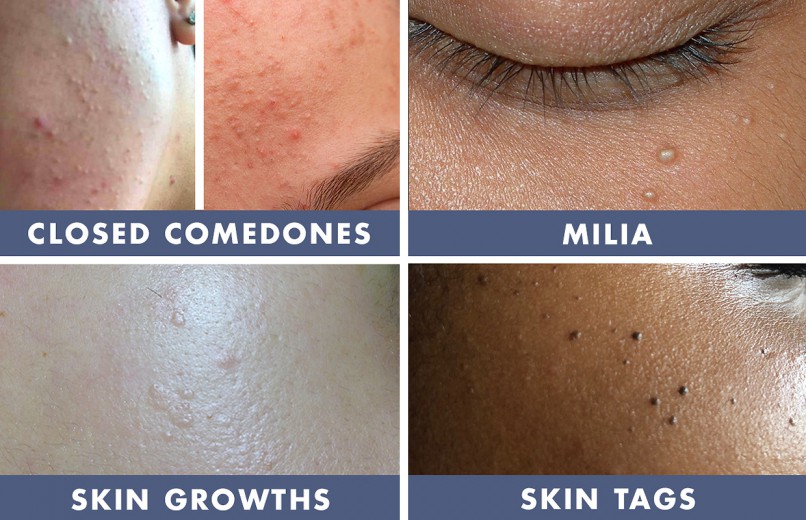
h) Skin tags characteristics & causes

These form a teardrop-shaped overgrowth of tissue which can be pigmented and which are often found on the neckline, collar, underarm or groin area. They are formed in areas of friction which is thought to stimulate the excess growth but which also causes discomfort and sometimes inflammation as the tag can pull or snag.
Skin tags will often present in groups although they can be singular.
Skin tags are common with virtually everyone – male and female – experiencing them at one time or another. They do however tend to appear more as we age or if we put on weight. They can also occur during pregnancy. Unlike warts, skin tags aren’t contagious and won’t spread to other others area of the body through touch.
The latest and most effective solutions for successful skin tag removal include short wave diathermy and Cryotherapy.
Short wave diathermy involves removing the skin tag with a small heated probe containing an electric current while cryotherapy utilises a freezing technique with liquid N2O using a Cryopen for the controlled destruction of unwanted skin tags. Both methods can be equally successful with the main difference being the way the skin reacts or heals.
Skin tags treated with SWD are likely to fall off during treatment. If this occurs, a small red lesion may be present at the site of the attachment. Tags that don’t fall off tend to drop off in a few days. In this case, the tag tends to shrivel with darkening of the area normal. Tags that don’t fall during treatment will do so in a few days. In either scenario, there should be little evidence a tag was ever-present after 4-6 weeks.
Cryotherapy treatment reacts differently. The skin tag will not come away during treatment. The nitrous oxide causes the death of the tissue which turns the lesion black. The lesion will drop off of its own accord within a few days or up to 4 weeks. In addition, larger skin tags treated with Cryotherapy may require a 2nd treatment. Short wave diathermy often requires only 1 treatment.
i) Milia characteristics & causes

Concealed very superficially under the skin, milia present themselves as small white plugs and often show as hard, solid lumps near the surface of the skin.
Milia is the retention of keratin and sebaceous material surrounding vellus hair. The exact cause is unknown although milia is often related to diet with a high cholesterol count, excessive vitamin C and too rich moisturising cream.
Milia can be treated with advanced electrolysis techniques using diathermy. This method gently dries them up so that the hard keratinised centre is broken down and will be absorbed by the skin following treatment. This is a much gentler way to treat them.
Removing milia with a microlance can damage the skin. Milia can appear between the eyelashes, on the eyelid itself, the cheek area or anywhere on the face or neck where dry skin is present. They tend to grow in size, become harder and then become noticeable to the eye and cosmetically unattractive. Some people may only suffer one and others exhibit a proliferation of up to 40 – 60 of them at any one time.
j) Seborrhoeic warts or keratosis characteristics & causes
Seborrhoeic warts or keratosis are very common harmless, often pigmented growths on the skin. They are benign growths due to a build-up of cells. They usually appear after the age of 40, although they can appear in younger people. The majority of older people will have a few seborrhoeic keratoses, while less will have large numbers. Seborrhoeic keratoses have a rough surface, and range in colour from golden brown to mid brown to almost black.

Seborrhoeic keratosis occurs most often on the trunk, but they are also common on the head and neck. Their numbers vary, and one person may have just one seborrhoeic keratosis whilst another can have hundreds. Once present, they usually stay, and new ones often appear over the years.
They can affect anyone, but on dark-skinned people, they can also appear as multiple small dark brown or black bumps, especially on the face and neck; in such a case this is called Dermatosis Papulosa Nigra.
Seborrhoeic keratosis is referred to as warts but they are not caused by the warts virus. They come with increasing age and are caused by an overproduction of keratinocytes. They are not malignant or contagious and can be treated.
k) What is cryotherapy and how is it used to treat blemishes?
The term ‘cryotherapy’ literally means ‘treatment using low temperature’ and refers to the removal of skin lesions by freezing them. The most common product used is liquid nitrogen. The Cryopen uses Nitrous Oxide.
l) Advantages and disadvantages of Cryopen
“The CryoPen uses a state-of-the-art, linear compression cooling technology that does not require handling of dangerous cryogenic gases and liquids, such as with existing, cryosurgical technologies of the 1970s. In addition to not being exposed to potentially dangerous gases or liquids, the pen-point precision and consistent freeze temperature of the CryoPen reduces the risks of serious burns.
The CryoPen also makes cryosurgery safer and easier for office staff, who will no longer have to regularly handle and order hazardous gases or liquids mandatory of most traditional methods. Plus, with the CryoPen, the treatment has minimal scarring and no need for anaesthetic.”
A wide variety of superficial benign and vascular lesions can be treated with Cryotherapy. These include but not limited to
- Hyperkeratotic lesions – skin tags, viral warts, verrucae.
- Vascular lesions – Campbell De Morgan spots
- Sebaceous lesions – Milia
- Pigmented lesions – Solar lentignes